Check out today’s Step 1 Qmax Question Challenge.
Know the answer? Post it below! Don’t forget to check back for an update with the correct answer and explanation (we’ll post it in the comments section below).
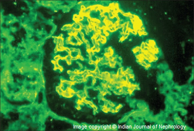 A 25-year-old previously healthy man comes to the emergency department with a 2-week history of persistent cough. He reports that he occasionally coughs up red, rusty sputum. Pulmonary function tests are notable for FVC of 60% and an FEV1:FVC ratio of 85%. A few weeks later, he presents to the emergency department complaining of abnormal urine color. A urinalysis is positive for hematuria. His temperature is 37°C (98.6°F), blood pressure is 150/95 mm Hg, and pulse is 105/min. Results of a renal biopsy are shown below.
Which of the following types of hypersensitivity reaction is this patient experiencing?
A. Type I hypersensitivity
B. Type II hypersensitivity
C. Type III hypersensitivity
D. Type IV hypersensitivity
———————–
Want to know the ‘bottom line?’ Purchase a USMLE-Rx Subscription and get many more features, more questions, and passages from First Aid for the USMLE Step 1, including images, references, and other facts relevant to this question.
This practice question is an actual question from the USMLE-Rx Step 1 test bank. For more USMLE Step 1 prep, subscribe to our Flash Facts and Step 1 Express video series. Score the best deal on all three products with USMLE-Rx 360 Step 1.


IgA nephropathy (Type II hypersensitivity)
It’s GPS (Type-2) anti-GBM antibodies
B)
It cannot be Berger’s even if you cannot interpret the biopsy picture. HSP associated with skin lesions.
By the way Berger’s is not a Type-2 it is a Type-3 HS. IgA-C3 deposits.
#@Hema S
b
type 2
goodpasture
Type 3 Goodpasture
GPS is Type II HSR.
Reference: Robbins.
The correct answer is B. This patient presents with a persistent cough, bleeding in the lungs, and blood in the sputum and urine. The renal biopsy shows a glomerulonephritis characterized by linear deposits of antibody along the glomerular basement membrane, indicating that this patient likely has Goodpasture syndrome, which is an autoimmune condition defined by the triad of glomerulonephritis (usually rapidly progressive or crescentic), pulmonary hemorrhage, and antiglomerular basement membrane (GBM) antibody formation. In this disease, there is a production of circulating autoantibodies, which are directed against an antigen intrinsic to the GBM, in response to an unknown inciting stimulus. The principal target for the anti-GBM antibodies is the ?-3 chain of type IV collagen, which is found in the basement membrane collagen.
The lungs and kidneys are affected because the vascular permeability in the alveoli and glomerulus leaves the basement membrane exposed, while it is covered in the rest of the body. It can also be referred to as anti-GBM antibody disease. This patient is experiencing hypertension, likely secondary to renal failure induced by the glomerulonephritis, as well as hematuria due to the glomerulonephritis. This is an example of a type II hypersensitivity reaction, in which antibody binds to antigen on a cell, leading to lysis by complement or phagocytosis. Because the damage in this disease is caused by the actions of antibodies on the GBM, diagnosis is made by immunofluorescence, which shows smooth linear staining along the basement membrane, like that shown in the question.
In type I hypersensitivity reactions, antigens cross-link preformed IgE on presensitized mast cells and basophils, which triggers the release of vasoactive amines. Examples of type I hypersensitivity reactions include asthma and anaphylaxis.
Type III hypersensitivity reactions are those involving immune complex deposition. Antigen-antibody complexes are deposited and activate complement. The activated complement attracts neutrophils, which release lysosomal enzymes. Serum sickness and the Arthus reaction are variants of the type III hypersensitivity reaction.
Type IV hypersensitivity reactions are delayed cell-mediated reactions. Sensitized T-lymphocytes encounter antigen and then release lymphokines, which leads to macrophage activation. Examples include skin tests for tuberculosis and transplant rejection.
A is not correct. In type I hypersensitivity reactions, antigens cross-link preformed IgE on presensitized mast cells and basophils, which triggers the release of vasoactive amines. Examples of type I hypersensitivity reactions include asthma and anaphylaxis. This patient’s presentation is not consistent with a type I hypersensitivity reaction.
C is not correct. Type III hypersensitivity reactions are those involving immune complex deposition. Antigen-antibody complexes are deposited and activate complement. The activated complement attracts neutrophils, which release lysosomal enzymes. Serum sickness and the Arthus reaction are variants of the type III hypersensitivity reaction. Other examples include rheumatoid arthritis and polyarteritis nodosa. This patient’s presentation is not consistent with a type III hypersensitivity reaction.
D is not correct. Type IV hypersensitivity reactions are delayed cell-mediated reactions. Sensitized T-lymphocytes encounter antigen and then release lymphokines, which leads to macrophage activation. Examples include skin tests for tuberculosis and transplant rejection. This patient’s presentation is not consistent with a type IV hypersensitivity reaction.
Thank you for the explanation, it’s very good to review material. Especially with very clear photos.
how to refill cbd catrirdge