It might not be the flashiest anatomical structure, but if you want to stand upright, and keep your retroperitoneal organs (like your kidneys) in place, the posterior abdominal wall is pretty important. Located at the back of the body, bounded by the lateral abdominal walls and the posterior parietal peritoneum, the posterior abdominal wall is a complex combination of muscles, bones, nerves, and vessels that provides structural support for the body and for the organs of the retroperitoneal space.
After listening to this AudioBrick, you should be able to:
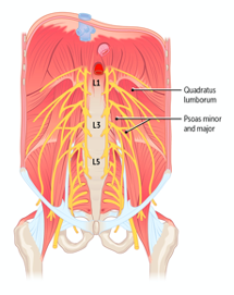
- Describe the structure and relationships of the posterior abdominal wall, including the aorta (including collateral channels), inferior vena cava (including collateral tributaries), lymphatics, muscles (psoas major, quadratus lumborum), sympathetic chain, and lumbar plexus.
- Describe the relationship of the kidneys in the retroperitoneal space as it relates to posterior abdominal wall anatomy.
If you haven’t subscribed to the Rx Bricks Podcast, we suggest you do it today!
Head to the homepage for the Rx Bricks Podcast to hear the full episode and subscribe so that you’re notified when the next one drops.


